What is Contrast-Induced Nephropathy?
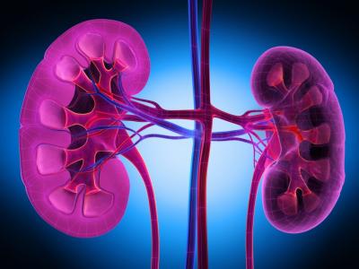
Have you ever heard of the term contrast-induced nephropathy (CIN)? This term may be unfamiliar for commoners, but this should not be the case for clinicians or health workers. We, especially clinicians and health workers, should be able to prevent contrast-induced nephropathy (CIN), and so we need to know what CIN is or need to recall how CIN occurs. Contrast-Induced Nephropathy is acute injury to kidney tissue caused by the entry of contrast agents into the body through the blood vessels. In normal kidney function, this damage is rare. However, in conditions of impaired kidney function (due to previous kidney disease), this contrast agent can exacerbate existing kidney damage. Contrast agents administered by intravascular injection, apart from having a toxic and apoptotic effect, may cause acute hemodynamic changes in the kidney which is characterized by an increase in renal vascular resistance and a decrease in the glomerular filtration rate. As a result, a significant decline in renal function can be expected to happen, especially in patients with pre-existing risk factors (eg diabetes, hypertension, congestive heart failure, and dehydration).
It should be noted, CIN is the third largest cause of acute renal failure in hospitals and contributes to 12% of cases of acute kidney injury (AKI). AKI itself is characterized by an increase in serum creatinine of more than 25% or >0.5 mg/dl from baseline within 48 hours. In an outline, the causes of AKI are divided into pre-renal, renal, and post-renal causes. The most common pre-renal causes are a lack of fluid supply to the kidneys such as dehydration, hypotension, embolism, and sepsis (severe infection). The post-renal causes that most often occurs is due to blockage by stones or mass pressure such as cervical cancer. While the most common renal causes are nephrotoxins, one of which is the contrast agent. Sometimes clinicians are faced with the unpleasant choice of having to use contrast even if the patient's kidney condition is poor.
In this modern and sophisticated era, medical technology is also moving forward as there has been sophisticated tools for imaging. In the process of establishing a diagnosis and medical intervention, we need tools that can reveal the flow of the blood vessels, digestive tract, and urinary tract before determining the disease and the subsequent actions. For example, to see tumors or infections in brain tissue and the lining of the brain, a computed tomography or CT-scan is needed. To obtain better results, CT-scan requires a contrast agent. This contrast agent aims to improve the visibility of areas, such as certain blood arteries, structures, or soft tissues, that appear faint in the image. Usually, a kidney function test is required before a patient has a CT scan with a contrast agent. The CT-scan can be done if their renal function is normal, as evidenced by normal blood urea and serum creatinine levels.
Peripheral angiography and intravenous pyelography are two other diagnostic procedures that make use of the contrast agents. Typically, peripheral angiography is performed to detect blood artery obstruction or blockage in the arms or legs. To determine whether the kidneys or urinary system are affected by disorders or obstructions, intravenous pyelography, or known as IVP, is utilized. The urinary tract stone is the most typical blockage. Both of these medical devices use contrast to reveal blood vessel and urinary tract flow using X-rays.
The most well-known and frequently performed medical intervention is percutaneous coronary intervention (PCI). PCI is used to place a stent in coronary heart disease. In an emergency, this interventional cardiac treatment becomes a priority with the relative temporary exclusion of the patient's renal function. In a study of the incidence of patients with renal failure due to diabetes, 50% were found to undergo cardiac angiography without the use of a low-osmolarity contrast agent and without adequate hydration.
Contrast agents have several types: a contrast agent that is inserted into the blood vessels and into the digestive tract (eaten or inserted through the anus), gadolinium-based intravenous contrast agent for MRI imaging and iodine for CT scan. The choice of these two ingredients is based on their properties that are easily cleared from the body in 24 hours through urine if the patient's kidney function is normal. In patients with renal impairment, deposition of contrast agent in skin tissue and other internal organs, including the brain, may occur. However, to date studies have found that even if the patient has end-stage chronic renal failure, the risk for this complication is relatively low. On the other hand, it becomes even more fatal for patients with previous impaired kidney organs. Contrast agents to reveal gastrointestinal tract are usually concentrated iodine or barium, both of which are not absorbed by the intestines and are excreted with the feces so they are not associated with kidney function.
In the last 10 years, contrast media such as Iopamidol, low osmolarity Iopromide (LOCM) and Iodixanol in the iso-osmolar form (IOCM) have been used to treat severe renal damage caused by contrast agents, in addition to adequate hydration and other professional preparations. Both can reduce renal vascular vasoconstriction. It should be noted that contrast agents can induce vasoconstriction or constriction of blood vessels and direct cell damage. The constriction of renal vasculature is related to the activation of the tubuloglomerular feedback (TGF) mechanism and the modulation of the production of intrarenal vasoactive mediators such as prostaglandins, nitric oxide, endothelin and adenosine. The TGF response itself depends on the type of osmolality of the contrast agent itself. Reduced synthesis of endogenous vasodilators (nitric oxide and prostaglandins) and increased synthesis of endogenous vasoconstrictors (Endothelin and adenosine) will increase the nephrotoxicity associated with these contrast agents. Renal function is further exacerbated by contrast agents by causing direct structural damage to the kidney cells. This damage process is associated with vacuolization of proximal tubular cells and necrosis to apoptosis of medullar ascending cells of the 'loop of Henle' in the kidney.
Thus, it is apparent that, in healthy kidney conditions, there will actually be no significant problems, because kidney function is supposed to get rid of toxic substances in the body. However, in conditions of kidney disorders, the use of contrast agents becomes a heavy burden for the kidneys. Research shows that the use of LOCM is 0.61 times safer in causing kidney damage compared to high osmolarity contrast agents.
Therefore, after realizing that there are risks in the use of contrast agents in daily medical practice, doctors and their team should pay more attention to kidney function before carrying out diagnostic procedures or medical interventions related to contrast substances. On the patient and family side, it would be better to know the risks of using the contrast agent and treat their kidneys with discipline in implementing a healthy lifestyle, including preventing diabetes and hypertension. This is all intended so that when there is a diagnostic procedure or action that requires contrast agent, the kidneys are able to completely clean the contrast agent (*).
Djoko Santoso
Professor Faculty of Medicine, Universitas Airlangga
Chairman of Health Department, Indonesian Council of Ulama, East Java